LONDON: The great thing about medical school cadavers is that they can’t die.
If a surgeon in training makes a mistake, there’s always next time. It is the last environment where medical errors have no consequences.


But 3D printing is changing that, giving even experienced operating room teams valuable practice on a model that looks and feels like the real thing. It has life-saving and life-altering implications.
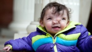
Violet Pietrok was born two years ago with a rare deformity called a Tessier cleft. The bones that normally join to form the fetal face had not fused properly.
As a result, Violet’s eyes were set so far apart, her vision was more like a bird’s than a human’s. She also had no cartilage in her nose.
But the corrective operation is extraordinarily complex. So Violet’s family turned to one of the world’s leading reconstructive surgeons, Dr. John Meara, at Boston Children’s Hospital.
Dr. John Meara has begun Violet’s series of surgeries. (Boston Children’s Hospital)
He warned them of the danger of making sophisticated cuts through the skull, very close to the optic nerve. “They might be very close to the brain,” Meara explained in an interview. “So the ability to make these cuts on the model first and see the trajectory of a sawblade or where that cut would come through in relationship to the eye is absolutely critical.”
To get that model, the simulation team at Boston Children’s took multiple MRIs of Violet’s skull and replicated it on a 3D printer.
It took more than a day to print, but the model is exact. Even the density of the bone is precise.