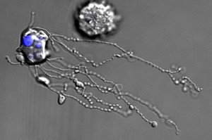

LONDON: In a first, researchers at La Trobe Institute of Molecular Science have managed to capture the complex stages of the death of a human white blood cell using time-lapse microscopy – something that has never been seen before.
Breakdown of dying cells, which was was once believed to be a rather random process, is effectively a high-regulated process say lead researchers, Dr Ivan Poon and Georgia Atkin-Smith.
Known for their role in defending human body against pathogens, the white blood cells are an innate part of our body’s immune system. Researchers, who observed the death of one of these cells, liken the even to fighter jet pilot ejecting from their downed aeroplane because certain molecules are pushed free from the dying cell, while others are left behind in the ‘wreckage’ of the cell fragments.
As seen in the video [embedded above], when the while blood cell starts to die it forms these lumps which push outwards and when the cell then explodes, it shoots out long ‘beaded’ protrusions which look like a necklace, which then breaks apart into individual ‘beads’.
Researchers say that proteins that are implicated in signal transfer, cell growth and maintenance all feature strongly in the beaded strings which are up to eight times longer than the host cell and that we have called ‘beaded apoptopodia’.
Post this explosion, the cells around the dead white blood cell can easily engulf these smaller pieces. Researchers believe that there could be certain molecules in the beads that could act as a warning system for other white blood cells signalling them to stay on the ‘Look out’ for any pathogens.

Tesla driverless system to use updated radar technology
WASHINGTON: Electric carmaker Tesla announced Sunday it was upgrading its Autopilot software to use more advanced radar technology. In a...