Customs Today
  • Home
  • Islamabad
  • Karachi
  • Lahore
  • National
  • Transfers and Postings
  • Chambers & Associations
  • Business
No Result
View All Result
Customs Today
  • Home
  • Islamabad
  • Karachi
  • Lahore
  • National
  • Transfers and Postings
  • Chambers & Associations
  • Business
No Result
View All Result
Customs Today
No Result
View All Result
Home Science & Technology Science

Powerful new microscope can help expose secrets of cancer, diabetes and multiple sclerosis

byCustoms Today Report
02/02/2015
in Science, Science & Technology
Share on FacebookShare on Twitter

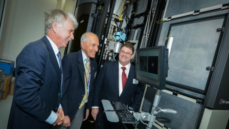
WASHINGTON: The $5m, three-metre tall microscope, unveiled at Monash University in Melbourne, allows researchers to see molecular structures at very high resolution.

“This is one of the most exciting days of my life,” said Professor James Whisstock, the Australian Research Centre’s director of advanced molecular imaging.

You might also like

Tesla driverless system to use updated radar technology

12/09/2016

Apple to develop its own self-driving technology

10/09/2016

Whisstock hopes the microscope will lead to better treatments for diseases such as cancer, diabetes and multiple sclerosis.

“Understanding our immune system is central to fighting cancer, infectious diseases such as malaria, and autoimmune diseases such as diabetes, rheumatism and multiple sclerosis.

“The key to understanding and treating these diseases lies in understanding how proteins and cells interact at the molecular level.”

Whisstock said the instrument highlights how physics and engineering together can be used to answer biological problems.

“We need physicists and engineers to be able to build these devices that can see the secrets of life,” he said.

Until now Australian scientists had to travel to Europe, Britain or the US to access similar microscopes.

“The problem with that is transporting biological material internationally is quite hard,” he said.

The Dutch-made Titan Krios instrument works by firing electrons through a sample.

Some of the electrons in the beam are deflected and these rays can be used to create a 2D image of the sample. Multiple 2D images can then be automatically pieced together to create 3D images of molecules.

Tags: Australian Research Centrediabetes and Multiple Sclerosisexpose secrets of cancerMonash University in MelbournePowerful new microscopeProfessor James Whisstock

Related Stories

Tesla driverless system to use updated radar technology

byCT Report
12/09/2016

WASHINGTON: Electric carmaker Tesla announced Sunday it was upgrading its Autopilot software to use more advanced radar technology. In a...

Apple to develop its own self-driving technology

byCT Report
10/09/2016

SAN FRANCISCO: Apple may not become an automaker, but it still wants to develop its own self-driving technology. The iPhone-maker's...

NASA spots slowest known magnetar

byCT Report
10/09/2016

WASHINGTON: Astronomers have found evidence of a magnetar - magnetised neutron star - that spins much slower than the slowest...

‘YouTubers’ outshining old-school television

byCT Report
09/08/2016

SAN FRANCISCO: A media revolution is taking place, and most people over 35 years of age aren’t tuned in. Millennial...

Next Post

European Space Agency finds how Philae lander dispatched to Comet 67P/Churyumov–Gerasimenko came to rest

  • Terms and Conditions
  • Disclaimer

© 2011 Customs Today -World's first newspaper on customs. Customs Today.

No Result
View All Result
  • Transfers and Postings
  • Latest News
  • Karachi
  • Islamabad
  • Lahore
  • National
  • Chambers & Associations
  • Business
  • About Us

© 2011 Customs Today -World's first newspaper on customs. Customs Today.