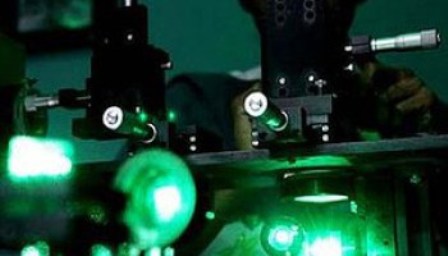
TORONTO: Researchers have taken live bacteria images using X-ray laser. This technique allows scientists to view inside the microbial organisms. These first-ever images taken of the inner workings of live bacteria were taken by researchers at the Linac Coherent Light Source (Lcls) at the Slac National Accelerator Laboratory, managed by the Department of Energy.
A fine aerosol of cells were exposed to pulses from an X-ray laser, and the electromagnetic energy was dispersed in patterns, which were recorded by a high-speed detector.


These high-energy X-rays quickly destroy living cells, limiting their use in medical and biological research. However, this new technique, known as “diffraction before destruction,” uses short bursts of electromagnetic radiation, making it able to record conditions within the cell before the target is destroyed. Synechococcus elongatus and Cyanobium gracile were both examined, using the Lcls.
This new study could pioneer new technologies that could image inner mechanisms of other bacteria, as well as viruses and other cells. Biological processes including photosynthesis and cell division could be witnessed live, potentially heralding new discoveries.
If the detector were better able to handle data collected in the experiment, it would have been possible to image the cells with a greater accuracy than what was accomplished. The images taken during the experiment were overexposed, in much the same way as a photograph taken in too much light. This problem could be corrected in future versions of the device, making it possible to see details in cells 20 times smaller than those seen in the current images.